Chinese and Australian researchers have developed groundbreaking imaging technology with the capabilities to transform the study of cellular processes and give insights into diseases like cancer, neurodegenerative disorders and metabolic conditions.
The study was conducted by researchers from the University of Sydney (UTS), Peking University and Eastern Institute of Technology, Ningbo.
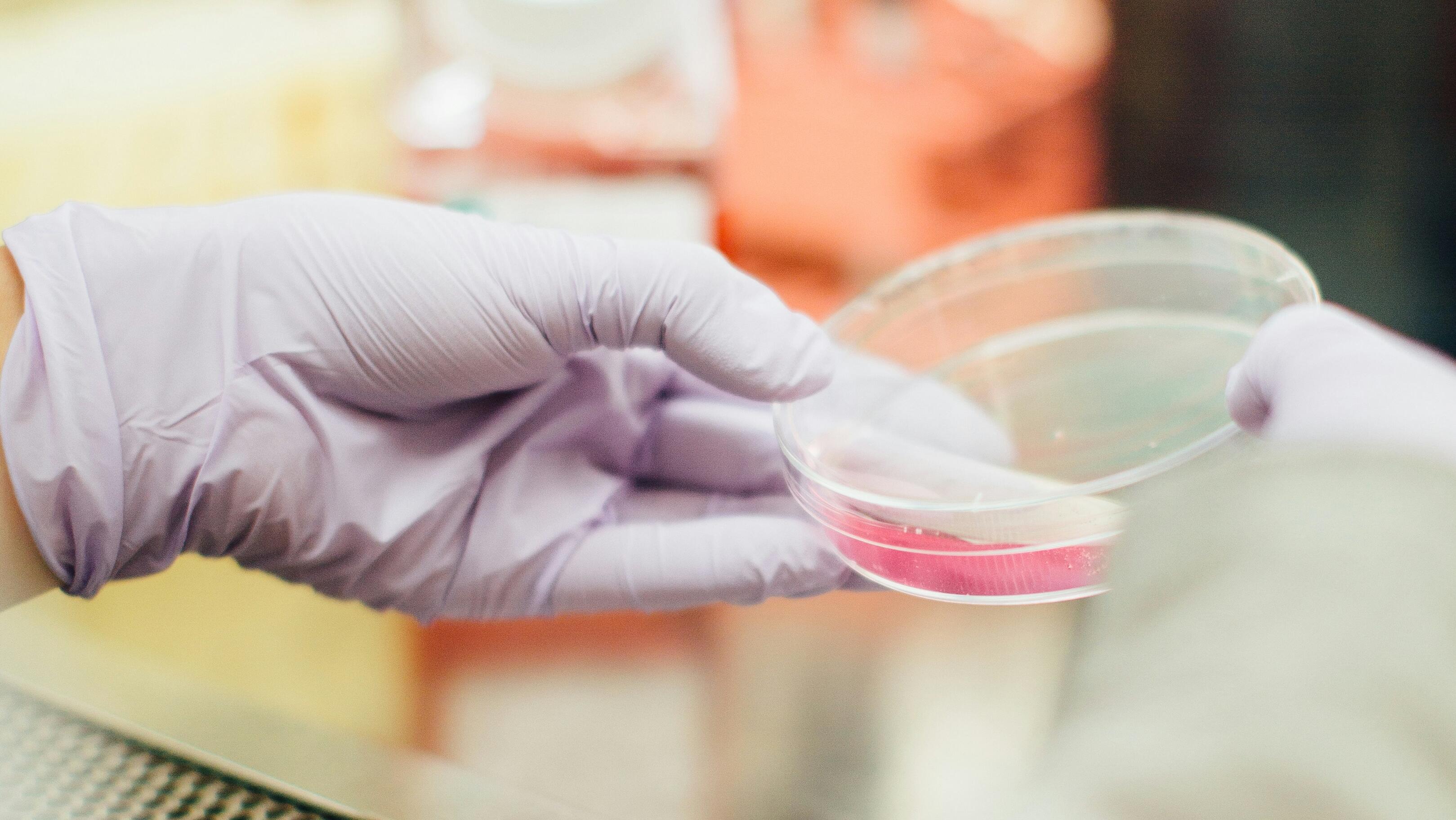
The research was recently published in Nature Communications and introduced an approach that combines super-resolution imaging with AI and deep learning to reveal subcellular structures and dynamics.
“This cutting-edge technology will open new doors in the quest to understand the intricate world within our cells," UTS professor and leader of the project, Jin Dayong said.
Dayong’s team is collaborating with medical research institutes, including scientists investigating cardiomyocytes, to advance heart disease research in hopes this breakthrough will lead to new insights and medical research advancements.
Dayong said the new method overcomes issues like face limitations in resolution that can cause phototoxicity and photobleaching, which damages cells through light exposure and colour restrictions found in traditional imaging tools.
This is through the use of a single laser and dye label that accurately predicts 15 different subcellular structures with just two detection channels, speeding up imaging and reducing cellular damage. It does this while producing high-resolution ‘optical fingerprints’ of organelles or cell compartments according to Dayong.
It is also highly adaptable and works across different microscopes, cell types and even complex living tissues to allow scientists to examine the 3D structure of live cells during cell division.